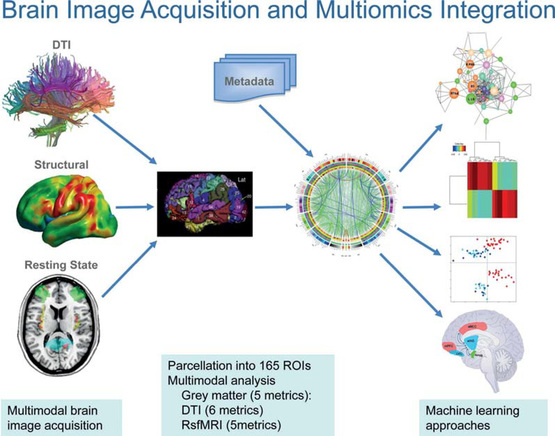
Figure 5.
Schematic of workflow from multimodal brain image acquisition to multiomics integration of brain and metadata. Acquisition of structural, anatomical (DTI), functional (resting state oscillations) and metabolic (MR spectroscopy, not shown) is followed by image processing and parcellation into multiple regions of interest (ROIs). These parcellated data undergo multiomics integration of different image modalities and clinical, behavioural and non-brain metadata using machine learning approaches. Such data-driven analysis approaches are expected to reveal distinct patters of brain-gut interactions. DTI, diffusion tensor imaging.

