Image 1.
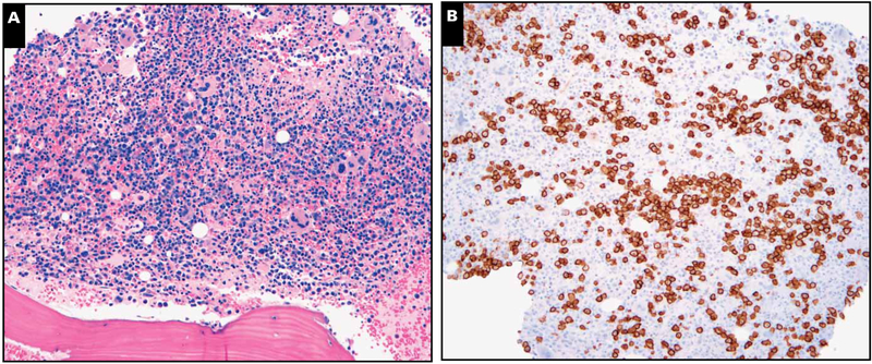
Markedly hypercellular marrow with plasmacytosis. A representative case with (A) marked marrow hypercellularity (H&E stain, magnification ×100) and (B) plasmacytosis (CD138 immunoperoxidase, magnification ×100) that was polytypic by light chain stains. This case did not show any lymphoid aggregates or stainable Kaposi sarcoma herpesvirus latency-associated nuclear antigen in the marrow biopsy specimen.

