Image 2.
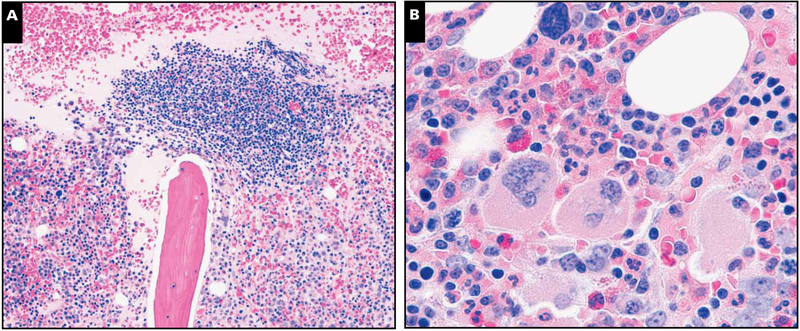
Illustration of a representative case with lymphoid aggregates and light chain–restricted plasma cells. A, Core biopsy specimen depicting paratrabecular lymphoid aggregate composed of lymphocytes, plasma, and few histiocytes with surrounding normocellular marrow with plasmacytosis (H&E stain, magnification ×40). B, Focal areas showed megakaryocytic clustering with mild fibrosis, but significant dyspoiesis was lacking in all 3 hematopoietic cell elements (H&E stain, magnification ×400).
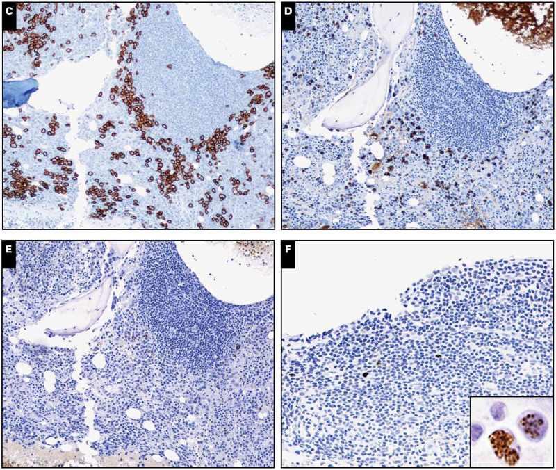
C, CD138 reveals marked interstitial plasmacytosis (magnification ×40). D, Plasma cells expressing monotypic λ light chain are seen rimming the edges of the lymphoid aggregate (magnification ×40). E, κ Light chain is largely negative (magnification ×40). F, Occasional lymphoid aggregates without well-formed germinal centers were noted to contain rare Kaposi sarcoma herpesvirus latency-associated nuclear antigen–positive intermediate-sized mononuclear cells with a characteristic stippled pattern of nuclear staining (inset magnification ×400). Most cells did not have the morphology of plasmablasts.


