Image 3.
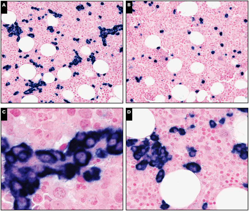
Case with “grape-like” clusters of light chain–restricted plasma cells. A, Clusters of atypical λ light chain–restricted plasma cells in an interstitial and perivascular location (in situ hybridization, magnification ×200). B, Corresponding κ light chain shows only singly scattered plasma cells (in situ hybridization, magnification ×200). C, D, High-power view depicting atypical perivascular and interstitial grape-like clusters of plasma cells possessing abnormal cytomorphology (in situ hybridization, magnification ×400). This case, however, was negative for Kaposi sarcoma herpesvirus (KSHV) latency-associated nuclear antigen and had no lymphoid aggregates. This patient died less than 1 year after KSHV–multicentric Castleman disease diagnosis with evidence of primary effusion lymphoma diagnosed at autopsy.

