Image 5.
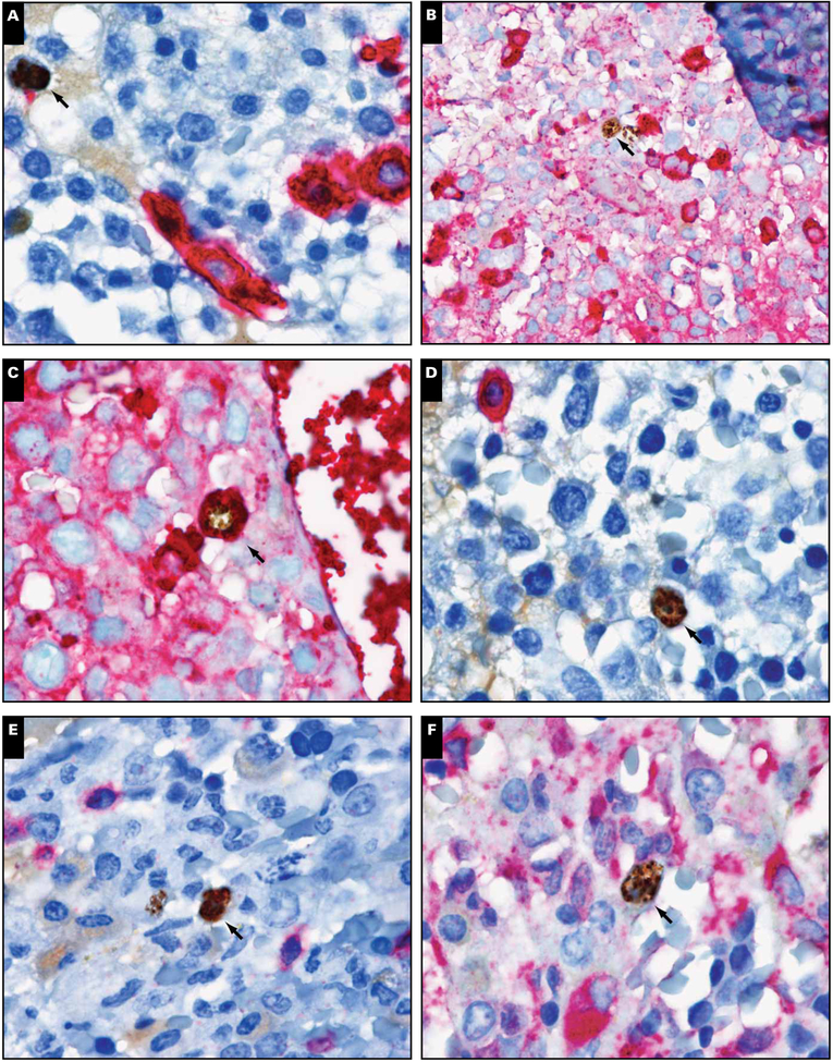
Immunohistochemical characterization of Kaposi sarcoma herpesvirus latency-associated nuclear antigen (LANA)–positive cells. Double staining of LANA-positive cells (brown; arrows) with (A) CD138, (B) κ, (C) λ, (D) CD20, (E) CD3, and (F) CD68 (all in red) revealed that LANA-positive cells stained only with λ (magnification ×1,000).

