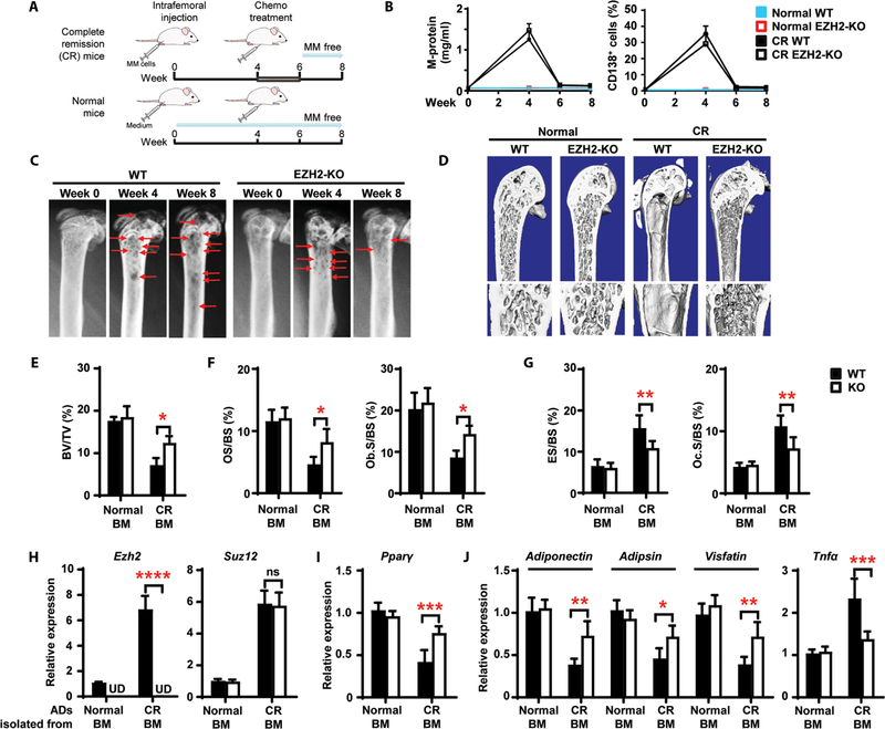
Fig. 7. Knockout of EZH2 in adipocytes heals resorbed bone in a mouse model of myeloma in remission.
Wild-type (WT) and adipocyte Ezh2-knockout (KO) mice were intrafemorally injected with the murine myeloma cell line Vk*MYC (1 × 10⁶ cells per mouse). After 4 weeks, bortezomib (1 mg/kg) and melphalan (2 mg/kg) were injected intraperitoneally into the mice thrice weekly for 2 weeks. Shown are the experimental schematic (A), the concentrations of M-protein in mouse sera, the percentages of marrow-infiltrated CD138+ myeloma cells (B), representative x-rays (C) of femurs from complete remission (CR) mice, and representative microcomputed tomography images of mouse femurs at week 8 (D). Red arrows, lytic lesions. (E to G) Percentages of BV/TV (E), OS/BS and Ob.S/BS (F), and ES/BS and Oc.S/BS (G) at week 8. (H to J) Relative mRNA expression for Ezh2 and Suz12 (H), Pparγ (I), and the adipokines Adiponectin, Adipsin, Visfatin, and Tnfα (J) in marrow adipocytes at week 8. Data are means ± SD (n = 5 mice per group, three replicate studies). UD, undetectable. *P < 0.05; **P < 0.01; ***P < 0.001; ****P < 0.0001. P values were determined using one-way ANOVA.

