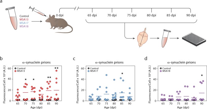
Fig 4. The rate of MSA prion propagation in TgM83+/- mice is variable.
TgM83+/- mice were inoculated with brain homogenate from a control (black) or MSA patient sample (MSA13 in red, MSA17 in blue, and MSA18 in purple). (a) Eight mice from each inoculation group were terminated every 5 days, starting from 65 days post inoculation (dpi) to 90 dpi. Frozen half-brains were homogenized and tested for α-synuclein prions using the α-syn140*A53T–YFP cell assay (× 103 A.U.). (b-d) None of the control-inoculated mice developed α-synuclein prions. The rate of α-synuclein prion formation varied across MSA-inoculated animals. * = P < 0.05; ** = P < 0.01.

