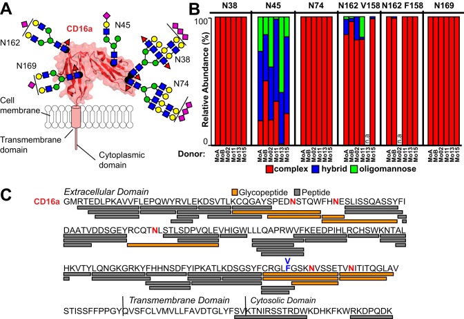
Fig. 2.
CD16a N-glycosylation composition varies by site and donor on monocytes. A, CD16a contains 5 N-glycosylation sites on the extracellular domain. These sites are modeled from the most abundant forms found on monocytes; the ribbon diagram and surface rendering are based on known structures of CD16a determined by X-ray crystallography. Carbohydrate residues are modeled as cartoons using the SNFG convention and roughly scaled to size (46). B, The relative abundance of each N-glycan type for the four individual donors and two donor pools as specified in Table I. n.a. - not applicable. C, ESI-MS/MS identified peptides and glycopeptides covering 84% of the CD16a sequence. Glycosylated Asn residues are shown in red. The V/F158 amino acid residue is labeled in blue.

