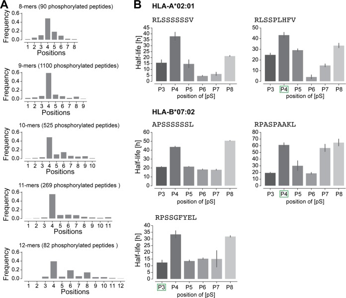
Fig. 3.
Phosphorylated positions in HLA-I ligands. A, Distribution of the position of phosphorylated residues in phosphorylated HLA-I ligands of lengths 8 to 12. B, Half-lives of HLA-I ligands for peptides with positions 3 to 8 substituted by phosphorylated serine. Green squares mark the position of the phosphosite (phosphorylated serine) for peptides found in MS data. Lack of green square indicates one unmodified peptide observed in MS data (APSSSSSSL) or one synthetic peptide (RLSSSSSSV) used in this in vitro assay.

