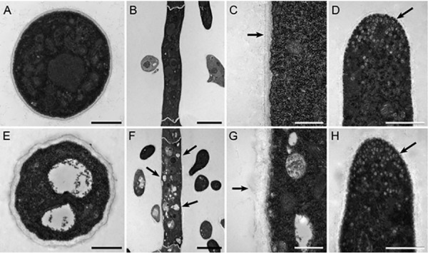
Fig. 9.

Transmission electron microscopy of thin sections of mycelia from R19 and transformant T625. A, Transverse cross section of R19 hypha. Bar is 1 μm. B, Longitudinal cross section of R19 hypha. Bar is 5 μm. C, Magnified longitudinal cross section of R19 hypha. Arrow points to cell wall boundary. Bar is 500 nm. D, R19 hyphal tip. Arrow points to small vesicles. Bar is 1 μm. E, Transverse cross section of T625 hypha. Note the two central hollow bodies compared with Fig. 9A. Bar is 1 μm. F, Longitudinal cross section of T625 hypha. Compared with Fig. 9B, arrows point to white enlarged endosomes. Bar is 5 μm. G, Magnified longitudinal cross section of T625 hypha. Arrow points to cell wall boundary. Note the thicker and bulbous cell wall compared with Fig. 9C. Bar is 500 nm. H, T625 hyphal tip. Arrow points to small vesicles as in Fig. 9D. Bar is 1 μm.
