Fig. 1-A Fig. 1-B.
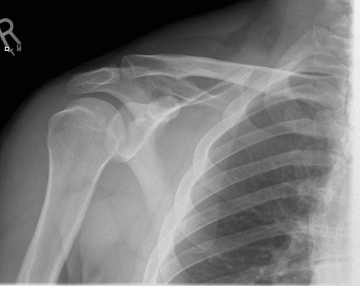
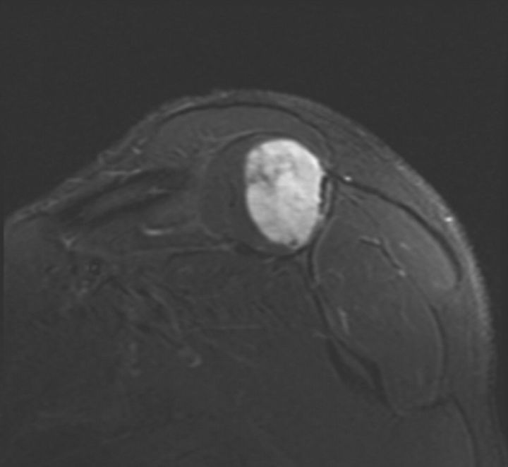
Fig. 1-A An anteroposterior radiograph of the right shoulder, showing a subtle destructive pattern within the scapular body. Fig. 1-B A coronal slice of a STIR sequence (short T1/Tau Inversion Recovery) magnetic resonance image of the shoulder, showing a partially enhancing solid mass within the belly of the right supraspinatus muscle. It was well circumscribed and measured 6.6 × 4.7 × 3.4 cm. The mass exhibits markedly high intensity on the STIR sequence.


