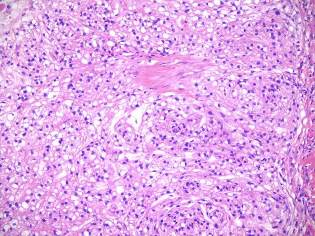
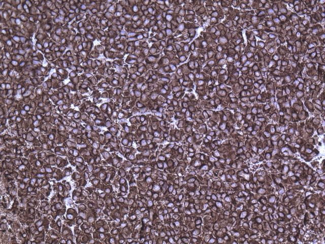
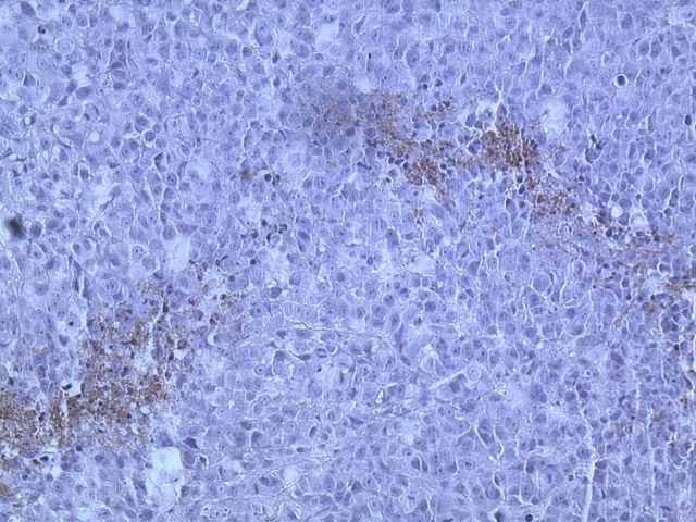
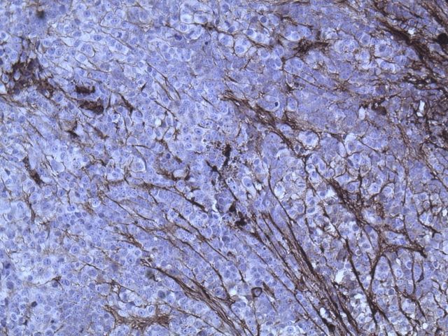
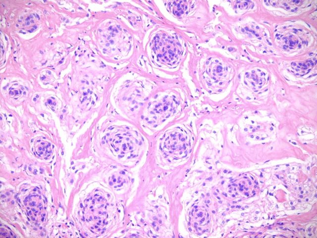
Fig. 2-A Fig. 2-B Fig. 2-C Fig. 2-D Fig. 2-E Fig. 2-F.
Hematoxylin and eosin staining of a tumor biopsy specimen (Fig. 2-A). Immunostaining for vimentin (Fig. 2-B), S-100 (Fig. 2-C), and cytokeratin AE1/AE3 (Fig. 2-D) (×10 for all). Hematoxylin and eosin staining of the resected tumor at 20× magnification (Fig. 2-E) and at 80× magnification (Fig. 2-F).

