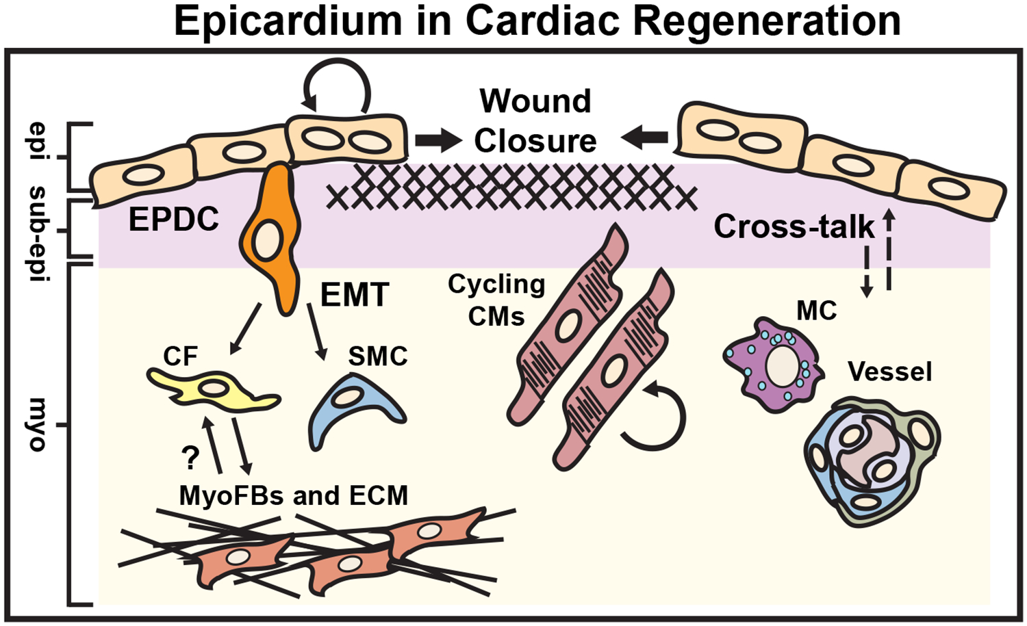
Figure 5. Epicardium in Cardiac Regeneration.

Cardiac regeneration occurs in response to a number of stimuli including apical resection, neonatal myocardial infarction, cryoablation or targeted ablation of the epicardium in both zebrafish and the early postnatal mammalian heart. The epicardium actively regenerates through the migration of “leader” binucleated epicardial cells initiating wound closure, and contributes to the formation of perivascular cells (smooth muscle cells or SMCs and fibroblasts or Fbs). Epicardium-derived fibroblasts have been shown to acquire myofibroblast phenotypes (MyoFb and ECM), which may be reversible during the resolution phase of the scar. The epicardium promotes macrophage (MC) recruitment, neovascularization as well as cardiomyocyte (CM) cell cycle re-entry via secretion of mitogens (dashed arrows). epi = epicardium, sub-epi = sub-epicardium, myo = myocardium. (Illustration credit: Ben Smith)
