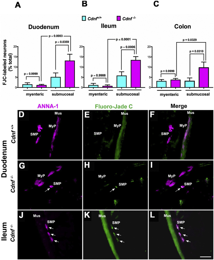
Fig. 4. Neurodegeneration selectively occurs in submucosal neurons of the duodenum, ileum and colon of Cdnf−/− mice.
Histochemistry was used with sections cut in a cryostat-microtome to analyze the proportions of neurons that were degenerating in the ENS of 1.5 month-old mice. Neurons were marked with ANNA-1 antibodies (magenta), degeneration with Fluoro-Jade C (FJC; green). The proportion of FJC-labeled neurons in the submucosal plexus of the duodenum (A), ileum (B) and colon (C) of Cdnf−/− mice (magenta bars) is significantly greater than that of their Cdnf+/+ littermates (turquoise bars). In contrast, the proportion of FJC-labeled neurons in the myenteric plexus of the duodenum (A), ileum (B) and colon (C) of Cdnf−/− mice does not differ significantly from that of their Cdnf+/+ littermates. [A, One way Anova, F (3, 34) =8.398 with Sidak’s multiple comparisons to compute the p values shown in the graph; B, One way Anova, F (3, 50) =21.93 with Sidak’s multiple comparisons to compute the p values shown in the graph; C, One way Anova, F (3, 92) =4.357 with Sidak’s multiple comparisons to compute the p values shown in the graph]. (D-L) Images of neurons (D, G, J) with coincident FJC labeling (E, H, K) in the duodenum and ileum of Cdnf+/+ (D, E, F) and Cdnf−/− (G-L) mice. Merged images are shown in panels F, I, L. Mus = smooth muscle layer; MyP = myenteric plexus; SMP = submucosal plexus. The arrows point to the locations of FJC-labeled degenerating neurons. The green fluorescence of the muscle layer is due to the non-specific FJC staining of smooth muscle. Scale bar, 35 μm.

