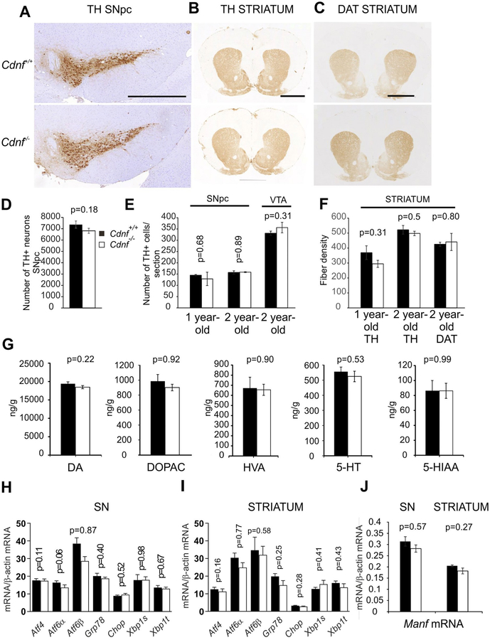
Fig. 6. Characterization of midbrain dopamine system in Cdnf−/− and Cdnf+/+ mice.
(A-C) Representative pictures of coronal sections from 2 year-old Cdnf+/+ (upper panel) and Cdnf−/− (lower panel) mouse brains immunostained with antibodies to TH and DAT at the level of the substantia nigra pars compacta (SNpc) and striatum. Scale bars: A = 100 μm, B-C = 200 μm. (D) Number of TH-positive neurons was quantified in the SNpc from Cdnf+/+ (n = 4) and Cdnf−/− (n = 7) 12 month-old male mice. (E) No differences were found between genotypes in the proportion of dopaminergic neurons quantified from scanned slides of TH-stained sections of SNpc of 9–12 month-old male mice (n = 2–3 mice/genotype) and SNpc and VTA from 2 year-old male mice (n = 3–4 mice/genotype) (F). TH-positive striatal fiber densities did not differ between Cdnf+/+ and Cdnf−/− mice quantified from 1 year-old mice (n = 2–3). No differences were found in striatal TH- and DAT-immunoreactive fiber densities in 2 year-old Cdnf+/+ and Cdnf−/− mice (n = 3–4) mice. (G) Dopamine (DA), DOPAC, HVA, serotonin (5-HT) and 5-HIAA concentrations were similar in striata from Cdnf+/+ and Cdnf−/− mice measured by HPLC from 9 to 12 month-old male mice (Cdnf+/+ mice n = 10, Cdnf−/− mice, n = 11, t-test; for DA and DOPAC, Mann Whitney U test). (H–I) qPCR analysis of transcripts encoding UPR-markers in SN (H) and striatum (I) of 9–12 month-old Cdnf+/+ (n = 3) and Cdnf−/− (n = 5) male mice. (J) qPCR analysis of transcripts encoding Manf in the SN and striatum of 9–12 months Cdnf+/+ (n = 6) and Cdnf−/− (n = 6) mice. Mean ± SEM, *p < .05.

