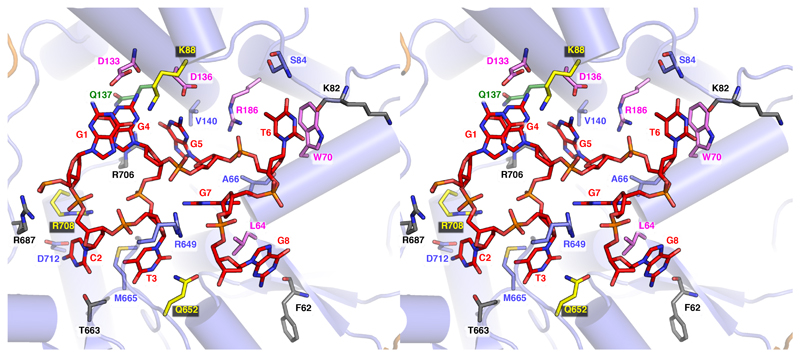
Fig. 3. Details of the full Chi-binding site.
A cross-eye stereo view of the Chi-binding site and interacting residues is shown. Interacting residues are shown as sticks and coloured according to previously published point mutation studies (19,20). Mutations at residues that failed to recognize Chi are in violet, those that showed altered/relaxed Chi recognition specificity are in green, and those that showed little or no influence after mutation are grey. Additional residues which failed to recognize Chi after mutation (22) are coloured in yellow.

