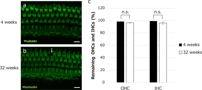
Fig. 4. IHCs and OHCs are similar in 4- and 32-week-old mice.
a, b Representative images of cochlear F-actin staining with phalloidin revealed no obvious differences in the number or morphology of OHCs and IHCs between mice at 4 and 32 weeks of age. The arrow in b indicates a loss of OHCs. c OHC and IHC survival was not significantly different between mice at 4 and 32 weeks of age. (n = 4 mice each.) n.s. not significant. Scale bars indicate 10 μm.

