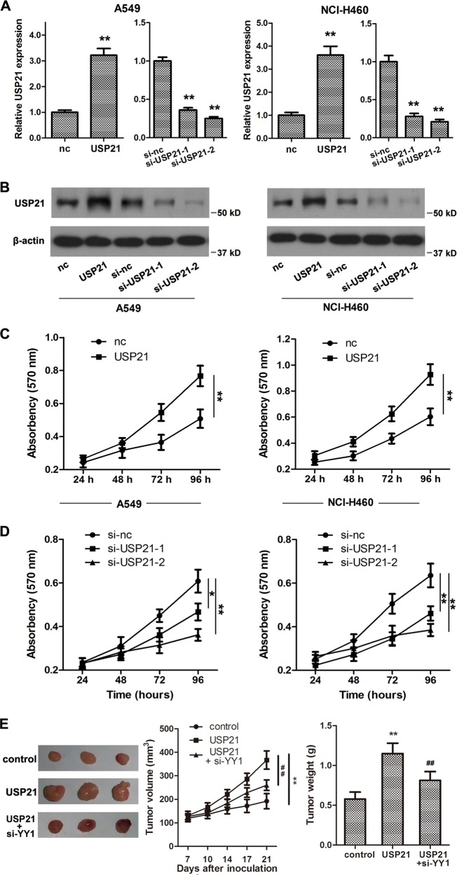
Fig. 2. USP21 promotes cell proliferation in vitro.
a Relative mRNA expression of USP21 after overexpression or silencing of USP21 compared with an empty vector control (nc) in A549 and NCI-H460 cells; n = 3. b Western blot analyses of USP21 after overexpression or silencing of USP21 compared with an empty vector control (nc). β-Actin was used as an internal control. c Cell proliferation was measured using an MTT assay at 24, 48, 72, and 96 h after transfection with the overexpression USP21 vector compared with the empty vector in A549 and NCI-H460 cells. d Cell proliferation was determined using the MTT assay at 24, 48, 72, and 96 h after silencing of USP21 or treating with the empty vector in A549 and NCI-H460 cells. e Representative images of tumor samples after 21 days of inoculation in nude mice models. Tumor weight was measured at the endpoint post 21 days, and the tumor volume was measured throughout the 21 days of inoculation. n = 5 mice per group in e. Each value is presented as the mean ± SD. n = 3. *p < 0.05, **p < 0.01 vs. the corresponding control; ##p < 0.01 vs. the USP21 group.

