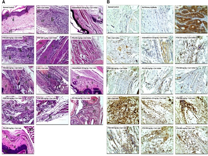
Fig. 3.
a Histopathological photomicrographs of paw tissues in normal control, initial and late sub-groups at 400 × total magnification. b Immunohistochemical photomicrographs of paw tissues in normal control, initial and late sub-groups at 400 × total magnification. ep epidermis, d dermis, ct connective tissues, li leukocyte infiltration, m muscle tissues, TTC tissue type control (colon cancer cells)

