Fig. 2.

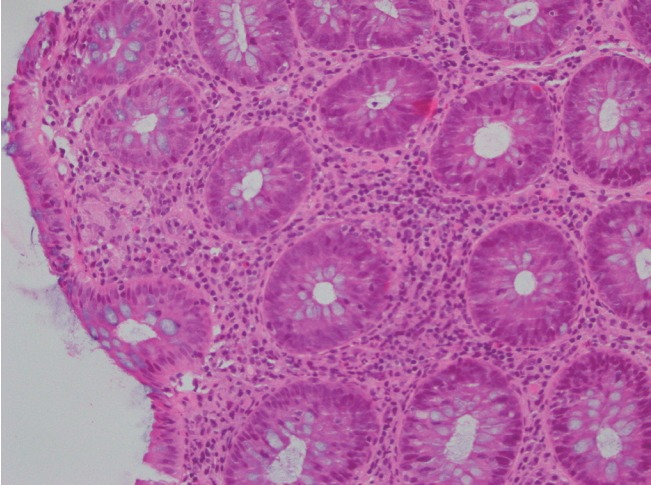
H&E stained slides from colonic biopsies (×200). The section shows mildly active colitis with increased mixed inflammatory cells in the lamina propria and an occasional cluster of histiocytes beneath the surface epithelium.

H&E stained slides from colonic biopsies (×200). The section shows mildly active colitis with increased mixed inflammatory cells in the lamina propria and an occasional cluster of histiocytes beneath the surface epithelium.