Figure 1.
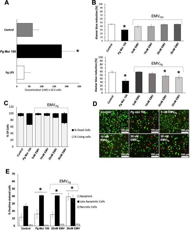
P. gingivalis promotes EMV shedding and alters endothelial cell viability. (A) The generation of EMV from naïve EC (control) and following 24 h of infection with P. gingivalis (Pg) (MOI = 100) or stimulation with Pg-LPS (1μg/ml) was measured in the supernatant by prothrombinase assay. Concentrations are represented by mean +/− SD from 3 independent experiments; *p < 0.05 vs control (unstimulated EC). (B) Metabolic activity of EC infected with Pg (MOI = 100) or exposed to EMVCtrl (upper panel) and EMVPg (lower panel) (5, 10, 20 and 30 nM) for 24 h measured by AlamarBlue assay. Results are expressed as mean +/− SD from 3 independent experiments; *p < 0.05 vs control (unstimulated EC). (C) Live-Dead assay to evaluate the ratio of live EC versus dead EC in cells exposed to P. gingivalis (Pg) (MOI:100) and EMVPg (5, 10, 20 and 30 nM) for 24 h. Results are expressed as percentage of live and dead EC (mean +/−SD). (D) Immunofluorescence imaging of live-dead staining (green: live EC, red: dead EC) for each condition after 24 hours of exposure. (E) Evaluation of type of cell death by flow cytometry after P. gingivalis infection and EMVPg (20 and 30 nM) exposure for 24 h. EC were labelled with Annexin-V FITC and propidium iodide (IPPE). All data were expressed as mean ± SD. *(p < 0.05 vs untreated cells).

