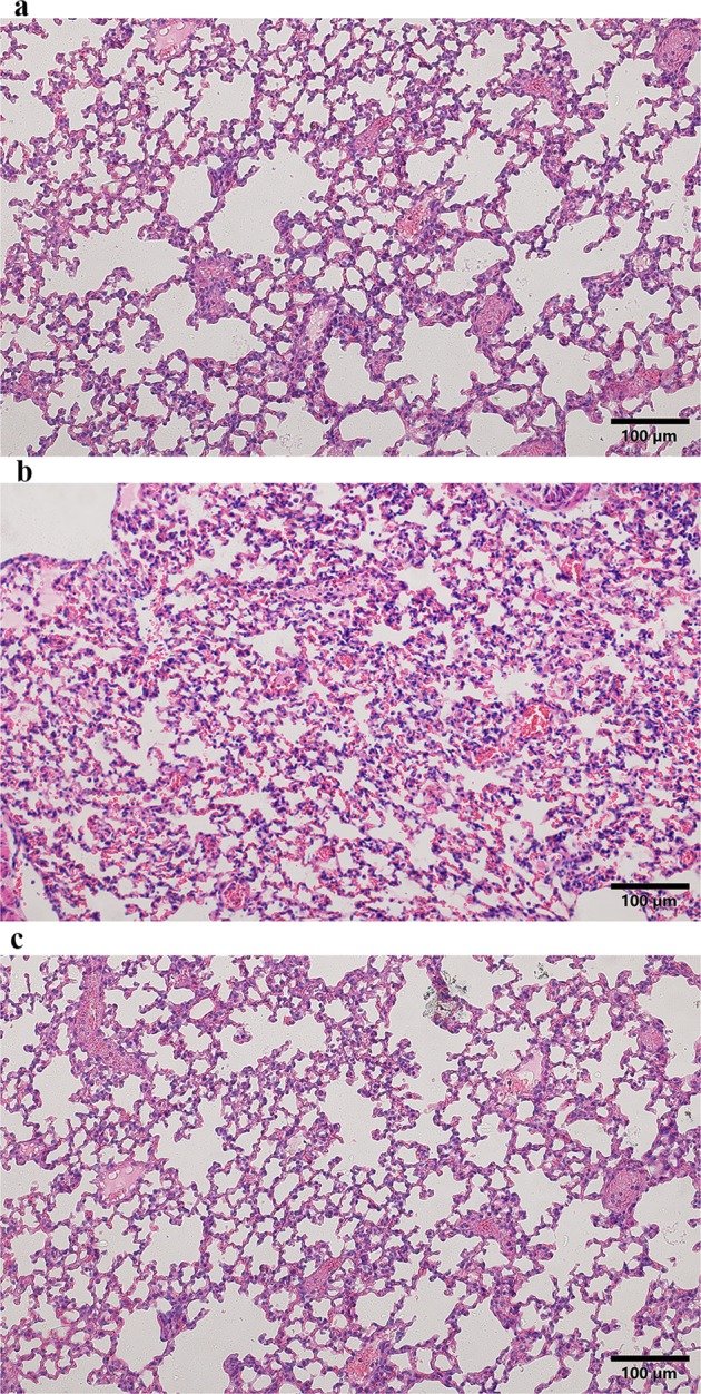
Figure 2.

Representative images of H&E staining of lung tissue in the different groups (magnification, 200×), (a) the control group, the alveolar structure of the control group was intact, and there was no edema in the alveolar wall and no inflammatory cell infiltration in the lung parenchyma; (b) the PQ group, a large number of inflammatory cells infiltrated, with obvious bleeding and clear membrane formation in the alveolar cavity; (c) the HMH group, The alveolar structure of the HMH group was slightly damaged with a small amount of inflammatory cells exudation and hemorrhage.
