Fig. 2.
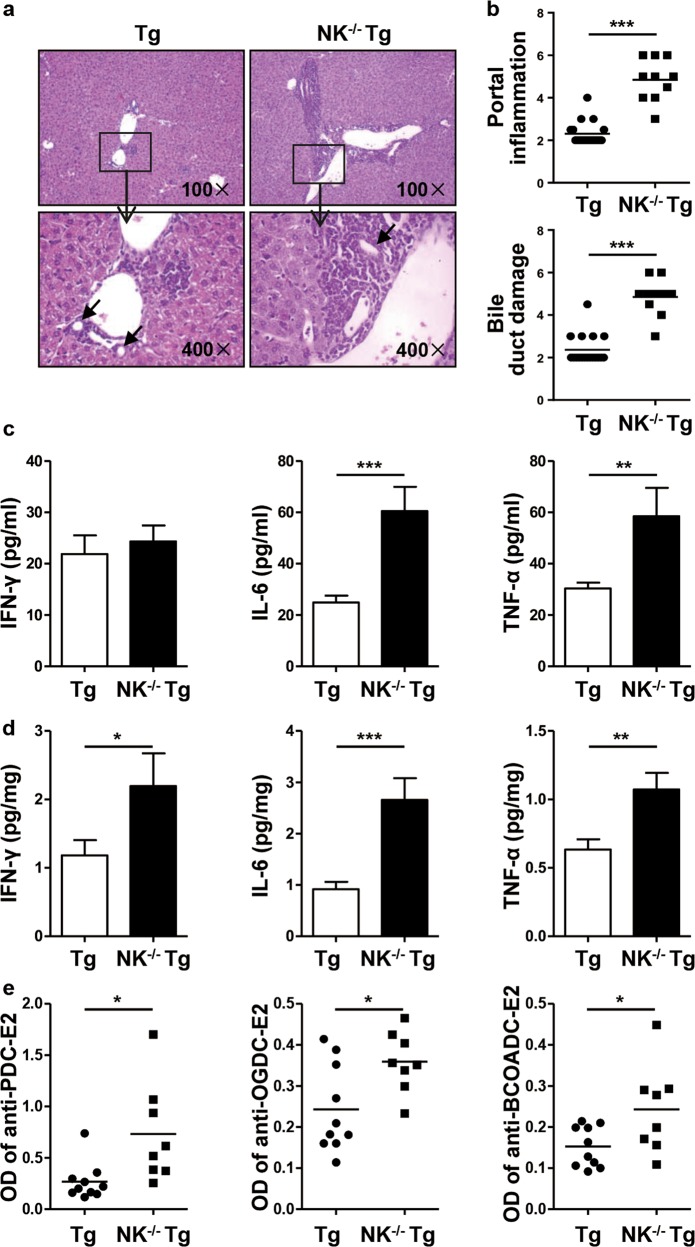
Autoimmune cholangitis was exacerbated in NK-/-dnTGFβRII mice compared to dnTGFβRII mice. a Representative H&E staining of liver sections from dnTGFβRII (Tg) and NK-/-dnTGFβRII mice (NK−/− Tg). The black arrows indicate the damaged bile duct. b Portal inflammation and bile duct damage scores in Tg mice (n = 18) and NK−/− Tg mice (n = 9). c-d The expression levels of IFN-γ, IL-6 and TNF-α in serum (c) and liver tissue homogenate (d) from Tg (n = 10) and NK−/− Tg mice (n = 6). e Serum levels of anti-PDC-E2, anti-OGDC-E2 and anti-BCOADC-E2 in Tg mice (n = 10) and NK−/− Tg mice (n = 8). *P < 0.05; **P < 0.01; ***P < 0.001

