Fig. 6.
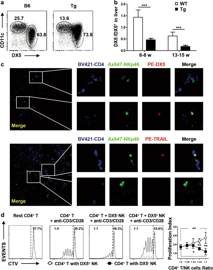
Liver-resident NK cells suppressed CD4+ T cell proliferation in vitro. a Hepatic NK cells were classified into DX5−CD11chi and DX5+CD11clo cell subsets by flow cytometry. b Representative ratio of DX5− to DX5+ NK cells in the early stage (n = 8) and typical symptom stage (n = 5–6) referenced in Fig. 6(a). c Representative immunostaining of frozen liver sections from Tg mice labeled with anti-CD4 BV421, anti-NKp46 Ax647, anti-DX5 PE and anti-TRAIL PE; confocal microscopy was then used to assess the location of NK cells and CD4+ T cells (200×). Here, NKp46+DX5− or NKp46+TRAIL+ represents liver-resident NK cells. d Representative FACS histograms of Cell Trace Violet-labeled conventional CD4+ T cells cocultured with hepatic DX5− or DX5+ NK cells from Tg mice under stimulation by 1 μg/ml anti-CD3 and 1 μg/ml anti-CD28. The data are representative of the results of three independent experiments. *P < 0.05; **P < 0.01; ***P < 0.001

