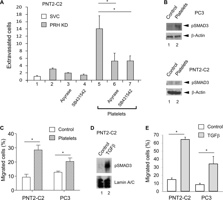
Fig. 2. ATP and TGFβ signalling increase prostate cell extravasation.
a PNT2-C2 cells expressing PRH shRNA (KD) or a scrambled vector control shRNA (SVC) were placed in a transwell chamber containing a confluent layer of HuVECs growing on Matrigel. Cells were pre-treated with platelets as in Fig. 1d in the absence and presence of apyrase treatment (columns 5 and 6) and in the absence and presence of 3 mm SB431542 (columns 5 and 7). After 24 h the number of extravasated cells was determined as described in Fig. 1b. Cells per field, n = 3 independent experiments, M + SD, *p < 0.01. b Immortalised prostate PNT2-C2 cells and prostate cancer PC3 cells were incubated with platelets (1:1) for 24 h. pSMAD3 levels were then determined using western blotting with β-Actin as loading control. c Cell migration over 18 h was then examined using a transwell chemotaxis assay. The percentage of cells migrated was determined by counting the cells on the top and bottom surfaces of the transwell filter using microscopy. Three independent experiments, M + SD, *p < 0.01. d PNT2-C2 cells were treated with 5 ng/ml TGFβ or vehicle for 48 h. Western blotting was then used to measure pSMAD3 levels using Lamin A/C as loading control. e PNT2-C2 cells and prostate cancer PC3 cells were treated with 5 ng/ml TGFβ for 48 h as above. Cell migration was then assayed as above. Three independent experiments, M + SD, *p < 0.01.

