Fig. 1.
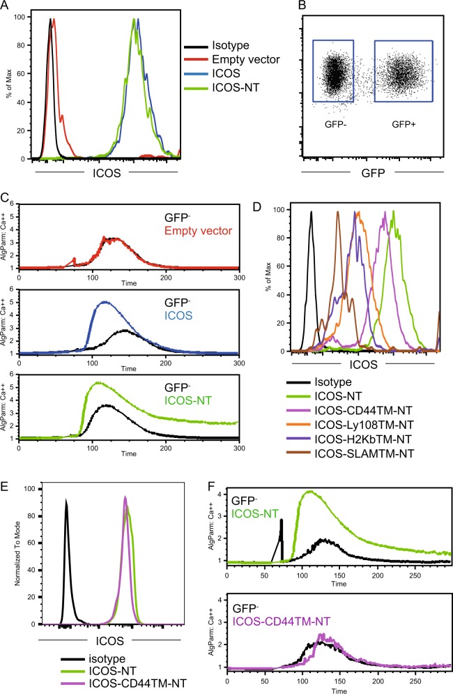
The ICOS transmembrane domain, but not the cytoplasmic domain, mediates the costimulation of calcium mobilization. a Surface ICOS staining of Jurkat cells lentivirally transduced with an empty vector or vectors expressing full-length ICOS or ICOS-NT. b Example of gating GFP+ stably transduced Jurkat cells and GFP− spike-in nontransduced control Jurkat cells for the calcium mobilization assay. c Calcium mobilization in Jurkat cells of the indicated types and the respective internal GFP- control cells after stimulation with biotinylated anti-CD3 (0.2 μg/ml) and anti-ICOS (2 μg/ml) antibodies coligated by streptavidin. The data represent the results of three independent experiments. d Surface ICOS staining of Jurkat cells lentivirally transduced with vectors expressing mutant ICOS molecules of the indicated types. e Surface ICOS staining after normalization of expression between ICOS-NT and ICOS-CD44TM-NT. f Calcium mobilization in Jurkat cells of the indicated types conducted as for c. The data represent the results of three independent experiments

