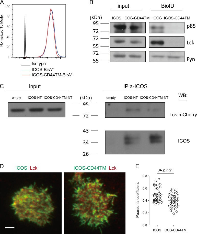
Fig. 4.
Transmembrane domain-dependent ICOS-Lck association. a Surface ICOS staining of Jurkat cells stably expressing BirA*-tagged ICOS or ICOS-CD44TM molecules. b Immunoblotting of p85, Lck, and Fyn in total lysates (input) or streptavidin-precipitated proteins from Jurkat cells that stably expressed BirA*-tagged ICOS or ICOS-CD44TM molecules after 24 h of culture in the presence of exogenous biotin. The data represent the results of four independent experiments. c Input (3% total lysates) or anti-ICOS immunoprecipitates of ICOS-NT or ICOS-CD44TM-NT Jurkat cells transduced with mCherry-tagged Lck were immunoblotted for p85 and ICOS. The data represent the results of three independent experiments. d Representative TIRF-plane images of EGFP-tagged ICOS or ICOS-CD44TM (green) and mCherry-tagged Lck (red) on Jurkat cells 10 min after being placed on anti-ICOS-incorporated planar lipid bilayers. e Pearson’s correlation coefficients for ICOS-Lck or ICOS-CD44TM-Lck colocalization on Jurkat cells. Each dot denotes one cell. The data presented were pooled from two independent experiments

