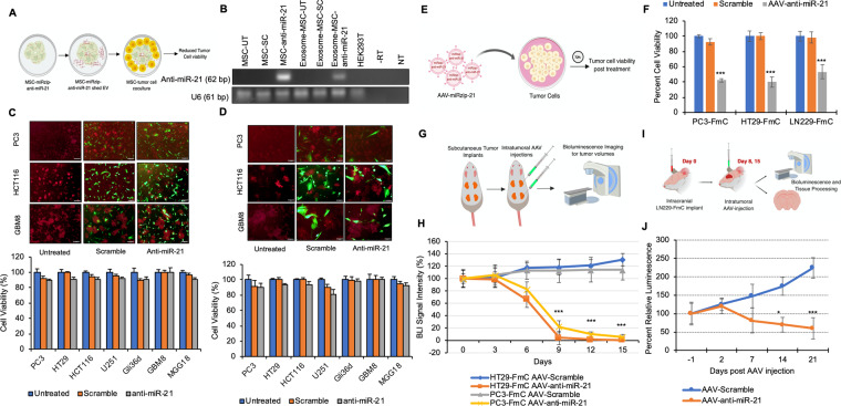
Figure 3.
AAV-miRzip-21 effectively reduces cancer cell progression in vivo (A) Illustration showing the proposed hypothesis of MSC based delivery of anti-miR-21 to tumor cells. (B) RT-PCR assay showing miR-21 expression in LV-miRzip-21 transduced-MSCs and exosomes extracted from transduced MSC. Negative and RT-minus controls are indicated by NT and -RT, respectively. UT and SC represent untreated and scramble control groups, respectively. (C) Plots and representative fluorescent micrographs of cancer cells cocultured with LV-miRzip-21 expressing MSCs at 3:1 ratio showing cell viability at 120 h. Scale bars: 100 uM (D) Plots and representative fluorescent micrographs of cancer cells cocultured with LV-miRzip-21 infected-MSCs at 1:1 ratio and corresponding cell viability at 120 h. Scale bars: 50 uM (E) Illustration showing the model for AAV transduction of tumor cells (F) Plot showing changes in tumor cell viability at 72 h post transduction with AAV-miRzip-21 and control. (G) Illustration of the in vivo subcutaneous model of colon and prostate cancers. (H) Plot showing changes in bioluminescence signal intensity overtime following AAV-miRzip-21 injection. (I) Illustration of the intracranial LN229-FmC animal model. (J) Plot showing changes in bioluminescence signal intensity overtime following AAV injection. Data presented as mean ± SD. ***(P < 0.01) and *(P < 0.5).

