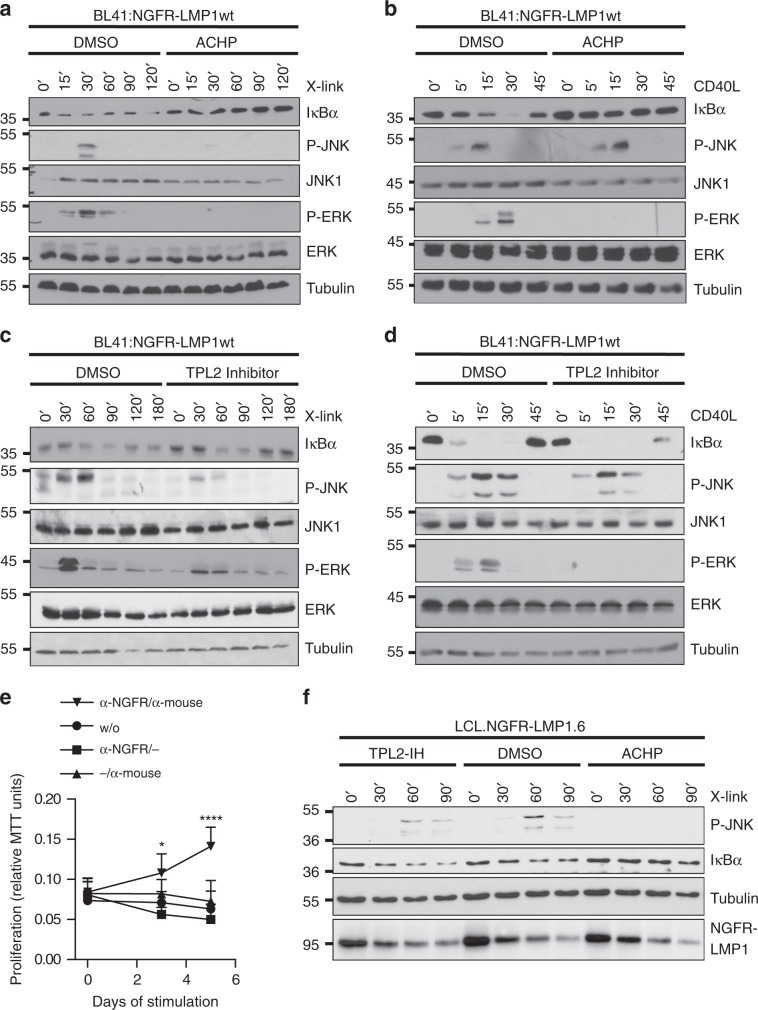
Fig. 6. LMP1 signaling to JNK differs from CD40 and requires IKK2 and TPL2 in B cells and LCLs.
a, b Effects of IKK inhibition on JNK, ERK, and canonical NF-κB signaling by LMP1 (a) and CD40 (b) in human B cells. BL41:NGFR-LMP1wt cells were treated with solvent (DMSO) or 5 µM ACHP. a NGFR-LMP1 activity was induced by antibody crosslinking (X-link), b CD40 was activated with 600 ng/ml soluble CD40 ligand (CD40L) for the indicated times. JNK, ERK, and NF-κB activation was detected in total cell lysates using the indicated antibodies. c, d Effects of TPL2 inhibition. Cells were treated with DMSO or 10 µM TC-S7006 (TPL2-IH). e Proliferation of LCL.NGFR-LMP1.6 cells depends on LMP1 activity. LCLs were established by infection of primary human B cells with EBV expressing NGFR-LMP1 instead of wildtype LMP1. Cells were deprived of crosslinking antibodies for 7 days to fully silence the chimeric receptor. Subsequently, the cells were re-stimulated as indicated and proliferation was measured as MTT conversion. Primary antibody: α-NGFR, secondary antibody: α-mouse IgG/IgM. The data are mean values ± SD of at least four biological replicates. f LMP1 signaling to JNK involves IKK2 and TPL2 in EBV-transformed human B-cells. LCL.NGFR-LMP1.6 cells were activated by antibody crosslinking in the presence of DMSO, ACHP, or TPL2-IH. JNK and canonical NF-κB activity was analysed by immunoblotting. NGFR-LMP1 was detected with the LMP1 antibody 1G6-3. e Statistical analysis was performed with two-way ANOVA at alpha 0.05, p-values: *p ≤ 0.05, ****p ≤ 0.0001. a–d, f The data are representative of at least two independent experiments. For immunoblot quantification and statistics see Supplementary Table 3.

