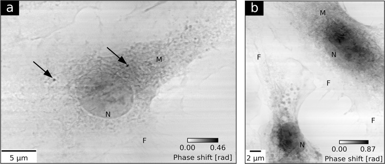
Figure 1.
Ptychographic imaging of two groups of macrophage cells. (a) presents the reconstructed phase of a macrophage treated with Fe2O3 nanocontainers targeting Mycobacterium tuberculosis. Black arrows indicate the internalization of two agglomerates of nanocontainers in the cell. (b) shows the ptychographic phase of two untreated control cells. Both images allow for identification of cellular nuclei (N), membrane ruffles (M) and filopodia (F).

