
Figure 2.
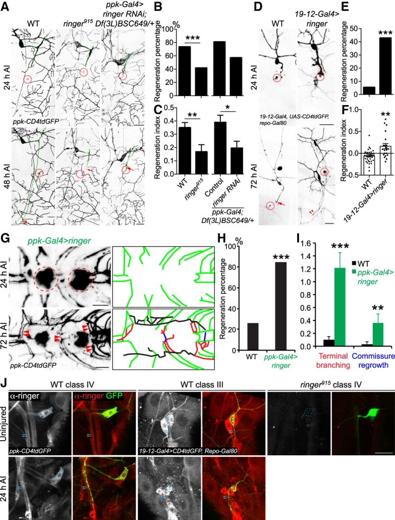
Loss-of-function and gain-of-function analyses for ringer in axon regeneration in the PNS and CNS. (A) C4da neuron axons robustly regenerate in WT. Ringer loss of function in a null allele, ringer915, significantly reduces the regenerative potential. C4da neuron-specific ringer RNAi in a sensitized background—deficiency Df(3L)BSC649/+ heterozygotes—also leads to reduced axon regeneration, indicating that ringer is cell-autonomously required for axon regeneration. The injury site is demarcated by the dashed circle. Arrow marks axon stalling while arrowheads show the regrowing axon tips. Axons are marked with dashed green lines. (B,C) Quantifications of C4da neuron axon regeneration with regeneration percentage (B) and regeneration index (C). N = 21 to 62 neurons from six to 16 larvae. (D) C3da neuron axons fail to regenerate in WT. C3da neuron-specific overexpression of ringer leads to increased axon regeneration. (E,F) Quantifications of C3da neuron axon regeneration. (G) C4da neuron specific overexpression of ringer increases axon regrowth in the VNC. The injury site is demarcated by the dashed circle. The regenerating axons are illustrated in schematic diagrams with terminal branching marked in red, commissure regrowth in blue, and other regrowing axons in black. (H) Compared with WT, which shows limited regrowth, the regeneration percentage is increased after ringer overexpression. n = 13 to 46 injured segments from seven to 29 larvae. (I) Ringer overexpression increases terminal branching and commissure regrowth of C4da neurons after injury in the VNC. N = 7 to 29 larvae. (J) Ringer expression pattern before and axon injury. Ringer protein is present in both C4da and C3da neurons and is detected in the axons before and after injury, as compared with the ringer915 negative control, which is devoid of ringer immunolabeling. Ringer is highly localized to the cell bodies and diffuse throughout the uninjured axon. After injury, ringer immunolabeling remains high in the soma and was concentrated within the proximal and medial portions of the injured axons. Strong immunolabeling of ringer protein is not observed in the growth cones or retraction bulbs of injured axons. The dashed circle marks the cell bodies and arrowheads mark the axons. (*) P < 0.05; (**) P < 0.01; (***) P < 0.001 by Fisher's exact test (B, E, H) or two-tailed unpaired Student's t-test (C, F, I). Scale bar, 20 μm.