
Figure 4.
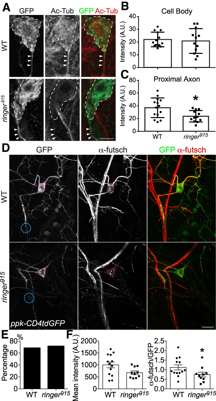
Ringer deficiency impairs microtubule integrity during axon regeneration. (A) Ac-Tub immunofluorescence, as viewed with confocal microscopy, demonstrates a reduction in microtubule acetylation in the cell body (dotted line) and axon (arrowhead) of injured ringer mutant axon. Scale bar, 5 µm. (B) There is no significant decrease in Ac-Tub immunofluorescence between WT (n = 13) and ringer mutants (n = 12), although variance is greater for ringer mutants. (C) Fluorescence intensity is significantly reduced from 37.02 ± 4.114 (n = 14) for WT to 23.73 ± 2.599 (n = 12) for ringer mutants within the proximal axon (5 µm). (D) In WT C4da neurons at 24 h AI, futsch is strongly expressed in both the cell body and the proximal and medial axon. Futsch immunolabeling is reduced in ringer915 mutant C4da neuron cell bodies and axons. The pink dashed circle marks the C4da neuron cell body and the teal dashed circle demarcated the injury site. Scale bar, 20 μm. (E) The percent of injured C4da neurons expressing futsch is comparable between WT and ringer mutants. (F) The fluorescence intensity of futsch staining in the soma of futsch-expressing C4da neurons (with and without normalization to GFP) is significantly reduced in ringer mutants. n = 10 to 13 neurons from five to seven larvae. (*) P < 0.05 by Fisher's exact test (E) or two-tailed unpaired Mann-Whitney test (B,C,F).