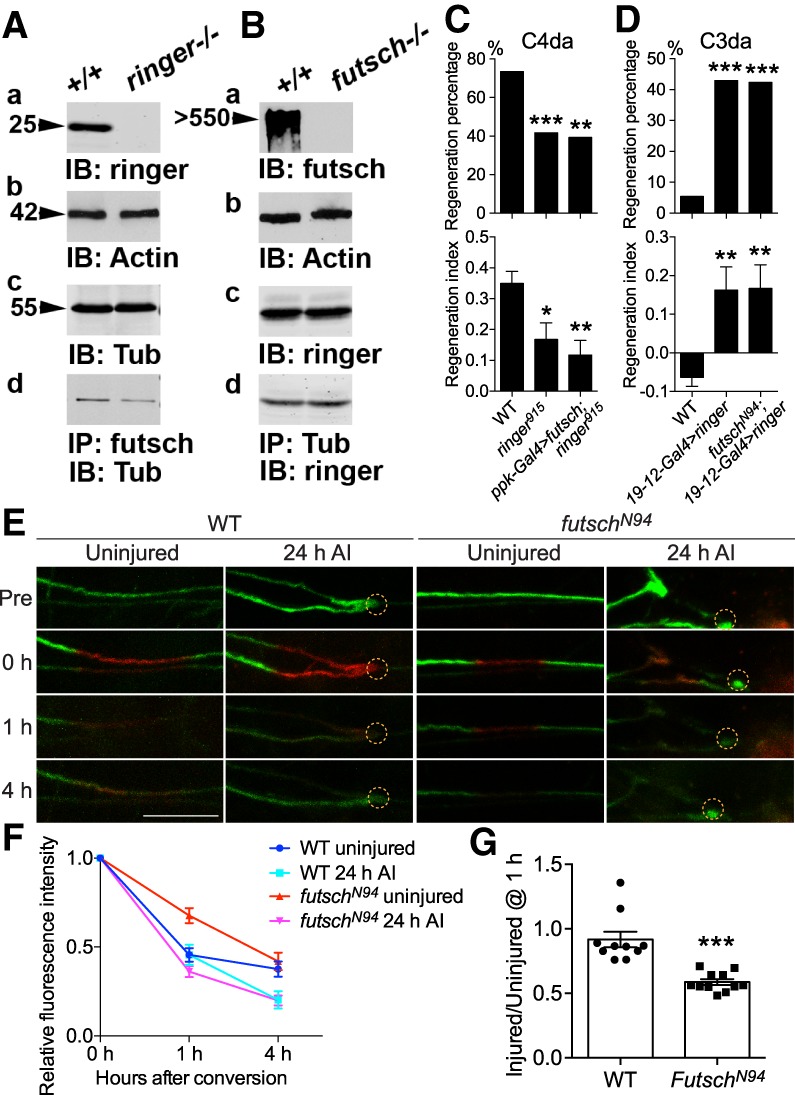
Figure 6.

Futsch binds to microtubule in a ringer-dependent manner and regulates microtubule dynamics. (A,B) Immunoblots from equal amounts of adult fly head lysates from WT and ringer mutants (A, panel a), and WT and futsch mutants (B, panel a) were immunoblotted for anti-ringer (A, panel a) and anti-futsch (B, panel a), respectively. Anti-Actin was used for loading control (A [panel b], B [panel b]). The levels of tubulin (A, panel c) and ringer (B, panel c) were unchanged in WT and ringer mutant lysates (A, panel c), and WT and futsch mutant lysates (B, panel c), respectively. IPs from the WT and ringer mutants (A, panel d), and WT and futsch mutant fly heads (B, panel d) show that while loss of any one of these proteins does not abolish the complex formation between the remaining two proteins, ringer is required to facilitate futsch binding to tubulin (A, panel d). (C,D) Quantifications of C4da (C) and C3da (D) neuron axon regeneration with regeneration percentage and regeneration index. N = 21–64 neurons from five to 16 larvae. C4da neuron overexpression of futsch does not rescue the reduced axon regeneration in ringer915 mutants, while futsch mutation does not suppress the enhanced axon regeneration induced by C3da neuron overexpression of ringer. (E–G) The assay for microtubule turnover/dynamics using tdEOS-labeled α-tubulin. A 20-μm region in the proximal axon was photoconverted by using a 405-nm laser. After 0, 1, and 4 h, the converted axon was checked for the remaining red tdEOS. Examples of photoconversion are shown in E. The dashed circle marks the injury site. Photoconverted segments were imaged at different time points and the red fluorescence was measured both in the conversion region and outside it. The difference between the signal in the conversion region and outside regions was normalized to the same measurement at 0 h. The relative fluorescence intensity (FI) over time is plotted (F). In WT, the relative FI is significantly reduced (*) in the injured versus the uninjured conditions at 4 h. At 1 h, the FI in uninjured futschN94 mutants is significantly higher (***) than that of WT, whereas the FI in injured futschN94 mutants is lower than the injured WT. (G) The ratio of the injured versus uninjured FI at 1 h was calculated and shows a significant reduction in futschN94 mutants. N = 7 to 11 neurons from five to 10 larvae. (*) P < 0.05; (**) P < 0.01; (***) P < 0.001 by Fisher's exact test (C,D, top), one-way ANOVA followed by Tukey's test (C,D, bottom), two-way ANOVA followed by Sidak's test (F), or two-tailed unpaired Student's t-test (G). Scale bar, 20 μm.
