
Figure 7.
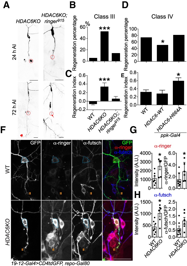
HDAC6 suppresses axon regeneration by inhibiting ringer and futsch. (A) Epistasis analysis of HDAC6 and ringer. Whereas HDAC6KO drastically promotes C3da neuron axon regeneration after peripheral injury, the enhancement is completely eliminated in HDAC6 and ringer double knockouts. The injury site is demarcated by the dashed circle. Arrow marks axon stalling while arrowheads show the regrowing axon tips. (B,C) Quantifications of C3da neuron axon regeneration with regeneration percentage (B) and regeneration index (C). n = 22 to 37 neurons from six to 10 larvae. (D,E) Quantifications of C4da neuron axon regeneration with regeneration percentage (D) and regeneration index (E). C4da neuron-specific overexpression of HDAC6-WT significantly reduces the regeneration percentage, without significantly affecting the regeneration index. On the other hand, overexpression of a dominant-negative HDAC6 mutant with the disrupted second catalytic domain HDAC6-H664A significantly increased the regeneration index. n = 24–64 neurons from six to 16 larvae. (F) Removal of HDAC6 drastically up-regulates ringer and futsch expression in both the soma and injured axon at 24 h AI, with a prominent increase within the growth cone. The dashed circle marks the cell body and arrowheads mark the growth cone. (G,H) Quantifications of the ringer (G) and futsch (H) immunofluorescence intensity in the soma (with and without normalization to GFP) shows significant increase in HDAC6KO, relative to WT. n = 6 to 7 neurons from three to four larvae. (*) P < 0.05; (***) P < 0.001 by Fisher's exact test (B,D), one-way ANOVA followed by Dunnett's test (C,E), or two-tailed unpaired Mann-Whitney test (G,H). Scale bar, 20 μm.