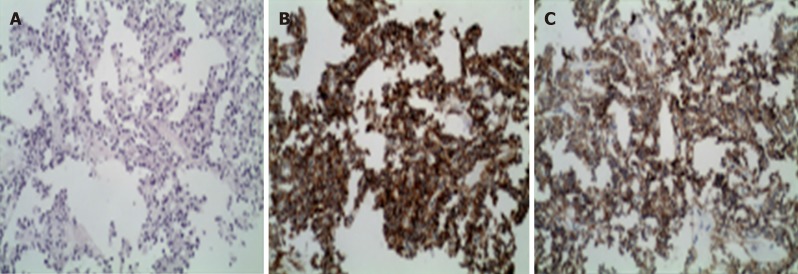
Figure 1.
Pathology of solid pseudo-papillary tumor of the pancreas with liver metastasis. A: The tumor is less heterogeneous, the cytoplasm is eosinophilic, and the tumor cells are arranged in a pseudo-nipple around the blood vessels; B: Positive cytoplasmic staining for CD10 (×200); C: Positive cytoplasmic staining for CD56 (×200).

