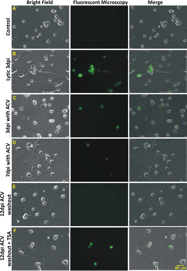
FIG 1.
Establishing a quiescent state of HSV-1 infection with ACV and triggering replication with TSA. Differentiated HD10.6 cells were infected with HSV-1, followed by fluorescence microscopy examining the progress of infection. (A) No-infection control. (B) Lytic infection. The virus replicated, and green fluorescence can be clearly seen at 3 dpi. (C) Infection with ACV at 3 dpi. Cells with green fluorescence can be observed with weaker intensity than that seen in lytic infection. (D) Infection with ACV at 7 dpi. Green fluorescent cells can barely be detected, and their number has decreased. (E) Infection at 12 dpi with 5 days of ACV washout. Fluorescence microscopy can hardly detect the green fluorescent cells, suggesting that a quiescent infection mimicking latency has been established and maintained. (F) Infection at 12 dpi with 3 days of ACV washout, followed by 2 days of TSA treatment. Fluorescence microscopy records more cells with stronger green fluorescence, suggesting that the dormant state of infection has been disrupted with increased viral gene expression and replication, a scenario similar to the reactivation observed in animal models.

