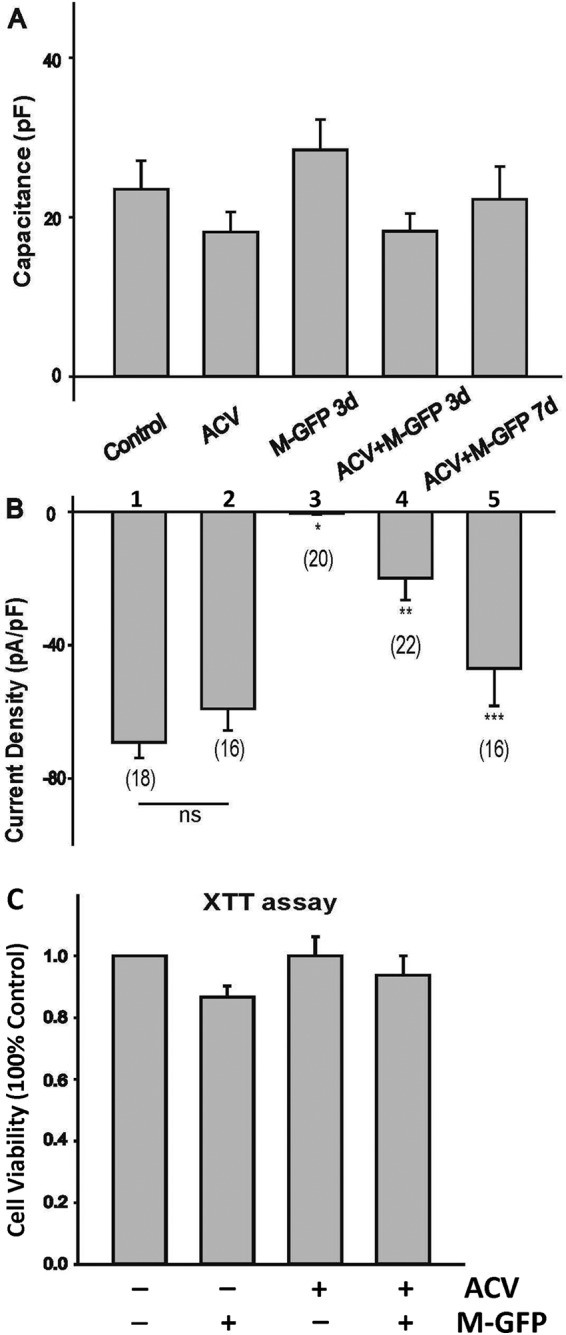
FIG 4.

Sodium current recording during HSV-1 latency establishment in HD10.6 cells. (A) Comparison of capacitances of HD10.6 cells under differentiation conditions. Control, no ACV treatment; ACV, ACV treatment; M-GFP 3d, lytic infection with GFP-labeled HSV-1 McKrae for 3 days; ACV+M-GFP 3d, latency establishment for 3 days; ACV+M-GFP 7d, latency establishment for 7 days. (B) Mean Na+ current densities generated in HD10.6 cells under different conditions. The voltage-gated Na+ current density was calculated by dividing the peak current amplitude by the cell capacitance. Asterisks indicate significant differences (P < 0.05) from the control group (*), the ACV treatment group (**), or the group infected with McKrae-GFP in the presence of ACV at 3 dpi (***). Numbers in parentheses indicate the number of cells recorded for each condition. ns, not statistically significant. (C) Cell viability was determined by the XTT assay. Four different groups were tested: control, M-GFP, ACV, and ACV+M-GFP. No statistically significant difference was detected among groups, although a minor reduction in viability at 3 dpi without ACV was observed.
