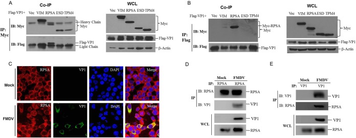
FIG 1.
FMDV VP1 interacted with host RPSA protein. (A and B) HEK-293T cells were cotransfected with 5 μg of Myc-vector-, Myc-VIM-, Myc-RPSA-, Myc-ESD-, or Myc-TPM4-expressing plasmids, and 5 μg of Flag-VP1-expressing plasmids for 36 h. The cells were then lysed and immunoprecipitated by anti-Myc antibody (A) or anti-Flag antibody (B). The immunoprecipitation (IP) complexes and whole-cell lysates (WCLs) were subjected to Western blotting using an anti-Myc, anti-Flag, or anti-β-actin antibody. (C) PK-15 cells were mock infected or infected with FMDV for 12 h, the subcellular localization of RPSA and FMDV VP1 was analyzed by immunofluorescence assay. Anti-VP1 (green) and anti-RPSA (red) antibodies and DAPI (blue) were used to stain the cells. (D and E) PK-15 cells were mock infected or infected with FMDV for 12 h. The cell lysates were then immunoprecipitated by anti-RPSA (D) or anti-VP1 (E) antibody. The IP complexes and WCLs were subjected to Western blotting with the anti-RPSA and anti-VP1 antibodies.

