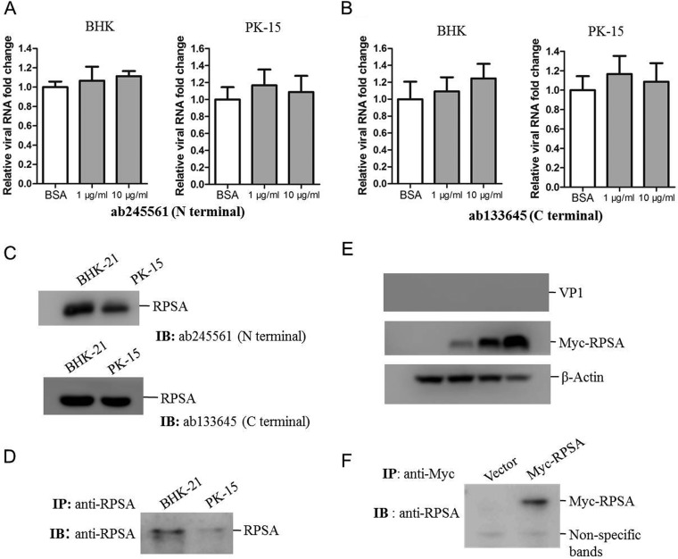
FIG 2.
RPSA was not responsible for FMDV entry. (A and B) BHK-21 and PK-15 cells were incubated with 10 μg/ml of BSA or 1 or 10 μg/ml of the anti-N terminus (A) or anti-C terminus (B) of RPSA antibodies for 1 h and then infected with FMDV (MOI of 0.5) for 12 h. The FMDV RNA levels were measured by qPCR. (C) BHK-21 and PK-15 cells were collected and lysed, respectively. The expression of RPSA was detected by using the two anti-RPSA antibodies. (D) The cell membrane proteins were extracted from the BHK-21 and PK-15 cells. The membrane fractions were then immunoprecipitated with anti-RPSA antibody and subjected to Western blotting. (E) HEK-293T cells were transfected with 0, 0.5, 1, or 2 μg of Myc-RPSA-expressing plasmids for 24 h. The cells were then infected with FMDV at an MOI of 1 for 12 h. The cell lysates were subjected to Western blotting with anti-VP1, anti-Myc, and anti-β-actin antibodies. (F) HEK-293T cells were transfected with 2 μg of vector or Myc-RPSA-expressing plasmids for 36 h. The cell membrane proteins were immunoprecipitated with anti-Myc antibody and subjected to Western blotting.

