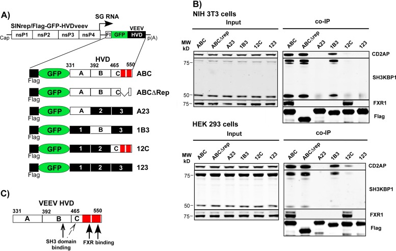
FIG 1.
VEEV nsP3 HVD interacts with murine and human FXR1 and SH3 domain-containing proteins via different fragments. (A) Schematic presentation of the designed SINV replicons and the encoded different fusions of Flag-GFP-HVD. Fragments with wt amino acid sequences are indicated by A, B, and C. The corresponding mutated fragments are labeled 1, 2, and 3, respectively. The elements of the C-terminal repeat in VEEV HVD are indicated by red boxes. Numbers indicate positions of amino acids in VEEV nsP3. Fl indicates position of Flag. (B) NIH 3T3 and HEK 293 cells were infected at an MOI of 20 inf.u/cell with viral particles containing SINV replicons encoding the indicated fusion proteins. Protein complexes were isolated using magnetic beads loaded with Flag-specific MAbs as described in Materials and Methods. Complexes were analyzed by Western blotting using the indicated primary and corresponding secondary Abs. Membranes were scanned on a Li-Cor imager. MW, molecular weight. (C) Schematic presentation of the positions of binding sites of the indicated host proteins in VEEV HVD.

