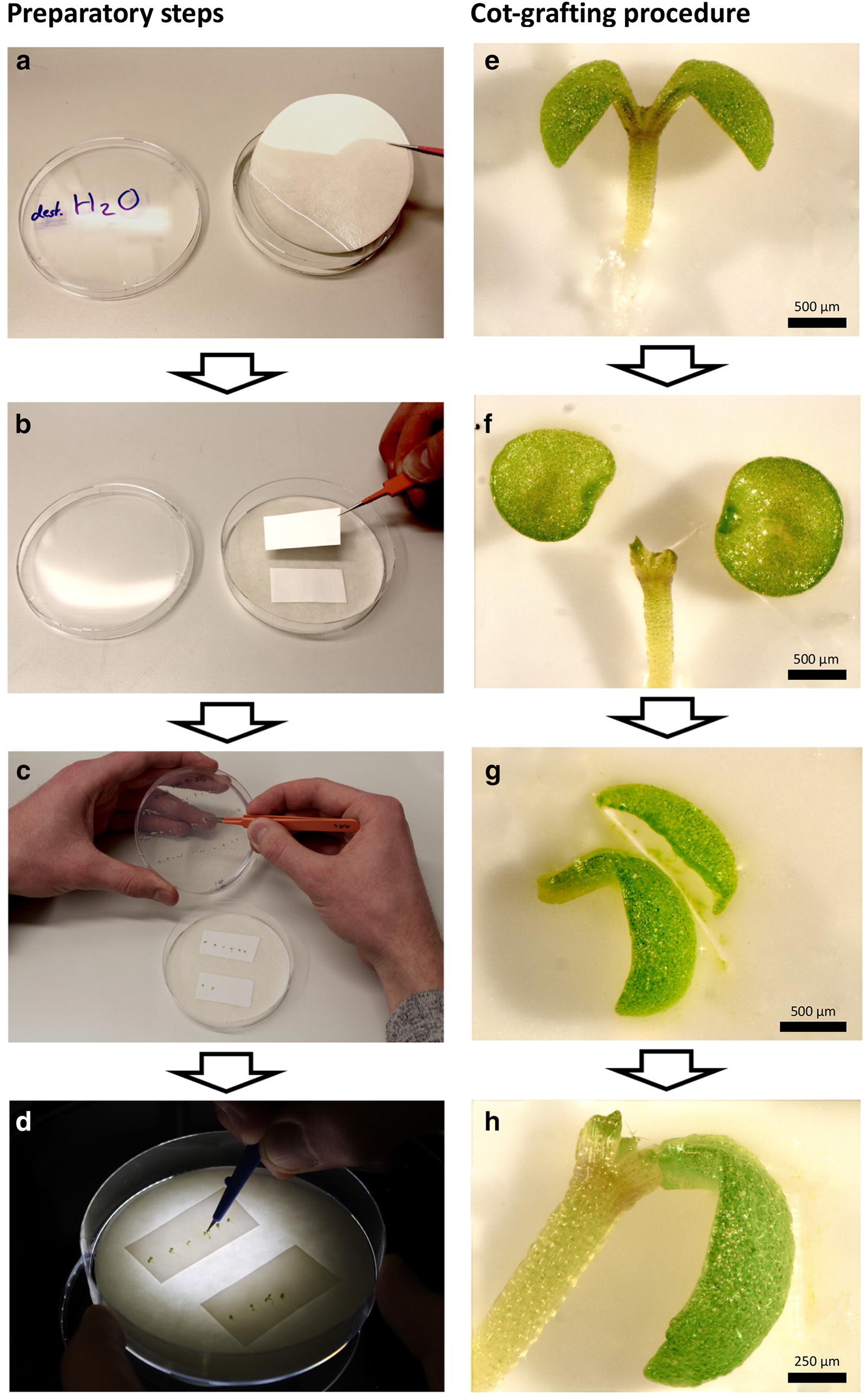
Fig. 1.

Workflow of cotyledon micrografting (cot-grafting) preparation and procedure. a–d Preparatory steps: Two layers of sterile filter paper were moistened with distilled water and placed into a sterile petri dish (a). Two stripes of nylon membrane were positioned on top of the filter papers (b). Vigorous seedlings were selected and placed onto the membrane using fine forceps (c). Cot-grafting was performed using a micro knife and a dissecting microscope (d). e–h Cot-grafting procedure: The cotyledons of donor and recipient plants were cut off (e, f). The donor cotyledon was cut alongside the central leaf vein on one side for optimal positioning (g). The donor cotyledon was finally transferred to the recipient plant (h). Scale bars correspond to 500 μm (e–g) and 250 μm (h), respectively
