Fig. 2.
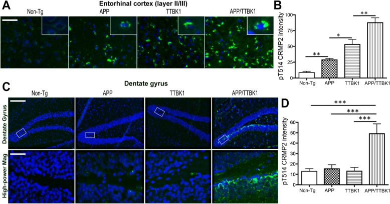
Transgenic expression of APP and TTBK1 synergistically enhance accumulation of pCRMP2 in the entorhinal cortex and dentate gyrus. APP (Tg2576), TTBK1, APP/TTBK1 and age-matched non-Tg mice at 10–11 months of age were subjected to immunofluorescence against pCRMP2 (T514, green) and Dapi (blue). a Layer II/III of the EC. Insets in A represent pCRMP2 accumulation in the neuronal cell soma. b Quantification of pCRMP2 intensity. The error bars indicate standard error of measurement (SEM). One-way ANOVA: F (3, 26) = 44.03 (p < 0.0001). c pCRMP2 staining was also observed in the subgranular zone and mossy fiber regions of the dentate gyrus of APP/TTBK1 mice but not in the other groups. Lower panel represents 20x original magnification of the subgranular zone. d Quantification of pCRMP2 intensity. The error bars indicate SEM. One-way ANOVA: F (3, 22) = 14.67 (p < 0.0001). b and d: *, **, and *** denotes p < 0.05, 0.01, or 0.001 as determined by one-way ANOVA and Tukey post hoc (N = 8 per group). Scale bars represent 50 μm (A), 200 μm (C, upper panel), and 30 μm (C, lower panel)

