Fig. 3.
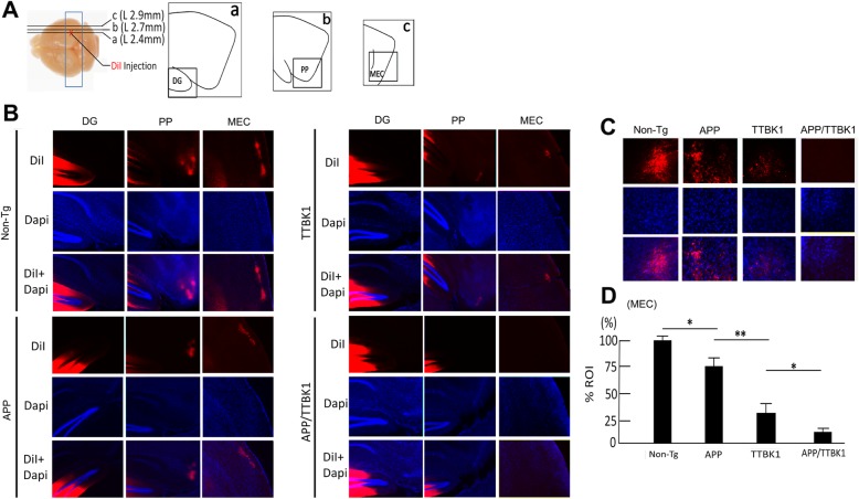
Transgenic expression of APP and TTBK1 induces axonal degeneration of EC neurons. a Stereotaxic DiI injection into stratum moleculare of dentate gyrus (DG) of a fixed and vibratome sectioned mouse brain. Transgenic littermates were sacrificed at 10 months of age and fixed brain tissues were injected with 0.5 μL of DiI. DG (a), PP (b) and MEC (c) regions were illustrated from the sections proximal to ML + 2.4 (a), + 2.7 (b) and + 2.9 (c). b DiI-labeled axon tracts (red) counterstained with Dapi (blue) in the DG, PP, and MEC in Non-Tg, APP, TTBK1, and APP/TTBK1 mice. c Retrograde tracing of DiI in the layer II/III region of EC of transgenic littermates. d DiI intensity measurements in the region of interest (ROI) of the layer II/III region of MEC. The error bars indicate SEM. * denotes p < 0.05 as determined by one-way ANOVA and Tukey post hoc (N = 6 per group). ANOVA F (3,20) = 82.48 (p < 0.0001)

