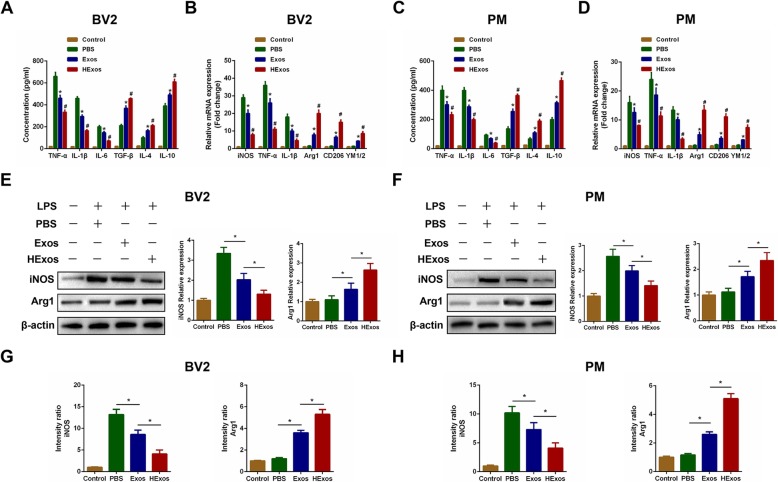
Fig. 4.
HExos shifted the microglia from M1 to M2 phenotype in BV2 and primary microglia in vitro. a The concentrations of pro-inflammatory and anti-inflammatory cytokines in BV2 microglia in the Control, PBS, Exos and HExos groups. b The mRNA expression levels of M1- and M2-related genes were detected by qRT-PCR in BV2 microglia in the Control, PBS, Exos and HExos groups. c The concentrations of pro-inflammatory and anti-inflammatory cytokines in primary microglia. d The mRNA expression levels of M1- and M2-related genes were detected by qRT-PCR in primary microglia. e and f The protein expression levels of M1- and M2-related genes were detected by western blot in BV2 and primary microglia in the different groups. g and h Quantification of the immunofluorescence intensity of iNOS and Arg1 from five different fields in each group. *P < 0.05 between the PBS and Exos groups, #P < 0.05 between the Exos and HExos groups

