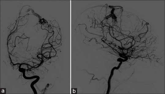
Figure 2.

Preoperative cerebral angiography. (a) Anterior-posterior view and (b) lateral view showed a nidus in the subcortical region of the left frontal lobe. It was supplied by distal branches of the left precentral artery and anterior parietal artery of the left middle cerebral artery. The venous drainage was to the cortical vein to the superior sagittal sinus
