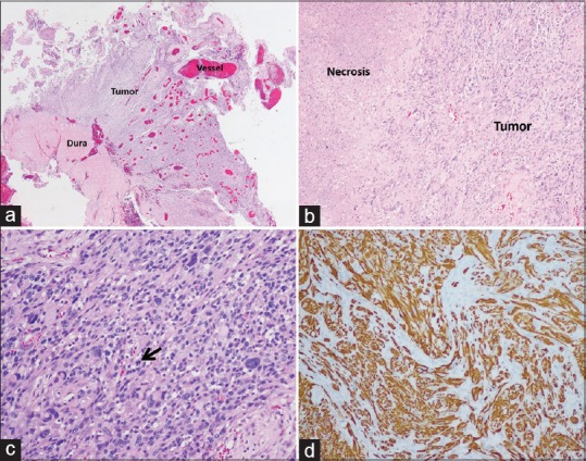
Figure 4.

Histological examination of the tumor. (a) Tumor attached normal dura matter and numerous blood vessels in varying sizes were within the tumor. (b) The necrotic area was observed. (c) The neoplastic glial cells had nuclear enlargement with hyperchromatic nuclei, and irregular nuclear membrane and mitotic activity could be observed (black arrow). (d) Immunoreactivity of the tumor was positive for GFAP
