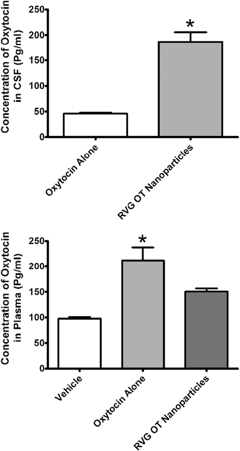
Fig. 4.
Concentration of oxytocin (OT) in cerebrospinal fluid (CSF) or plasma 2 h after intranasal administration of vehicle, OT alone, or OT encapsulated in rabies virus glycoprotein (RVG)-coated nanoparticles. Levels of OT in CSF following vehicle administration are not shown as they were below the limit of detection. TOP: Concentration of OT in the CSF. BOTTOM: Concentration of OT in the CSF. OT levels were determined in CSF and plasma by ELISA. Abscissae: Treatment condition. Ordinates: Concentration of OT in CSF (top) or plasma (bottom). All assessments were conducted in duplicate. * = p < 0.05 as assessed by unpaired t-test.

