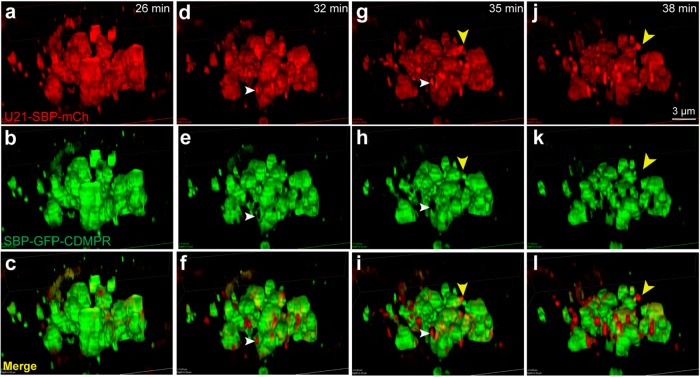
FIGURE 4:
U21 segregates from CDMPR in the Golgi. Live imaging of RUSH in HeLa cells expressing RUSH U21 (U21-SBP-mCh) and RUSH CDMPR (SBP-GFP-CDMPR). Reporter molecules were released from their hooks with biotin and superresolution imaging was performed at 3-min intervals after both proteins had reached the Golgi. Still images were extracted from the movie at the indicated times after biotin addition. (c, f, i, l) Merged images. White arrowheads in d–i show U21-SBP-mCherry segregating from SBP-GFP-CDMPR in the Golgi (d, g) and absence of SBP-GFP-CDMPR from sites of U21-SBP-mCherry concentration (e, h). (f, i) Merged images. Yellow arrowhead (g–l) shows a budding U21-containing carrier devoid of SBP-GFP-CDMPR. Scale bar: 3 μm.

