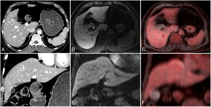
Figure 5:
65-year-old male status post abdominal-peritoneal resection with a nonspecific lesion on CT (A and D). At hepatobiliary phase MRI (B and E) and FDG PET (C and F), no suspicious features are identified. This case demonstrates how PET/MRI provides improved characterizations of liver lesions compared to PET/CT.

