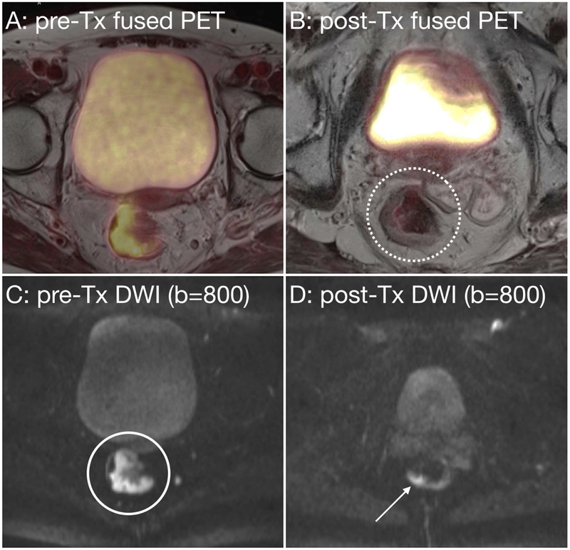
Figure 6:
72-year-old female with rectal adenocarcinoma before (A) and after chemoradiation (B). Post-therapy images suggest residual tumor on T2 weighted images, given that there was only a 24% reduction in tumor volume (B, white dotted circle). FDG-PET shows a marked reduction in metabolic activity, with a reduction of the SUVmax from 13.7 to 4.5 (a 67% reduction). DWI images show a decrease in tumor that demonstrates restricted diffusion (C, white circle and D, white arrow). MRI indicated partial response on T2 weighted imaging, while PET imaging suggests potentially a complete response. At pathology this was confirmed a complete response.

